
随着结构心脏病学再次登上心脏病治疗的舞台,作为第三只眼的超声心动,成为了患者筛选、策略制定、手术指导以及随访必不可少的工具。不少心脏内、外科的医师开始恶补超声心动图的相关技能。但是和世界上任何一个其他国家都不同,在中国由于缺少心脏专科医师的培训,大多数临床心内科医生和超声心动严重脱节,对超声心动图的进展所知甚少。其实随着心脏病学科的整体发展、超声技术的进步,超声心动也丰富了许多非常重要的信息,对临床有更强的指导意义。而临床医生如果能掌握这些新的技能,也许如同添上双翼,更加深入、自由地管理复杂的心血管疾病。
在最新一期的美国心血管病杂志JACC上,JACC imaging 的主编明尼苏达大学的Y. Chandrashekhar教授,发表主编社论梳理了2018-2019年,有关于超声心动的最新热点。
本期结构Vision 将随着这篇文章的步伐,来看看心内科医生必须要了解的超声心动方面的新进展。
判断左心功能不全,射血分数(LVEF) 就足够吗?
无疑,超声心动图的重要作用之一是对心力衰竭及其相关的综合征进行诊断及评估。射血分数(LVEF)也是评价左心室功能最重要的指标。但EF值有时并不可靠,在测量准确性方面容易存在谬误,对心衰患者的预后及心脏功能恢复的预测能力方面也有限。心室整体纵向应变( GLS)是近10余年来关注的热点,也仍然是2019年度超声心动最受关注的领域。大量证据表明在对心力衰竭的预后预测方面,GLS优于传统方法如射血分数(EF)。
那么什么是GLS 呢?那么GLS测量为什么优于LVEF值呢?
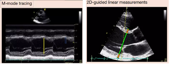
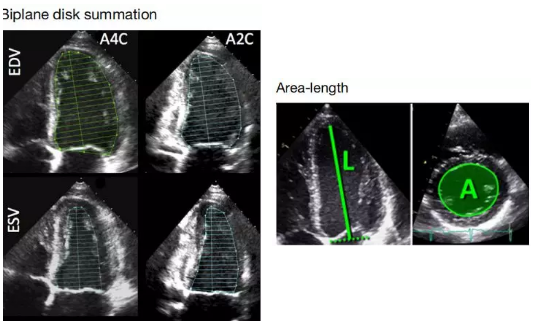
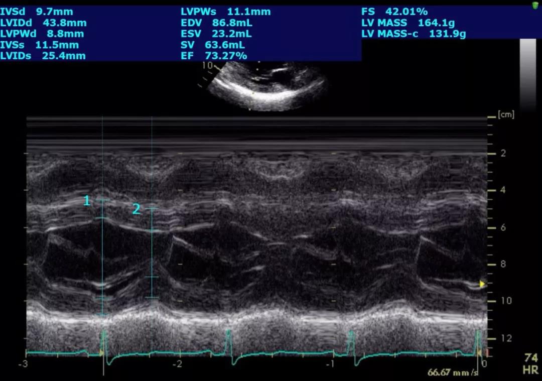
我们传统上测量EF值的方法,最常应用的也是指南上推荐的方法——Simpson 法,是指左室舒张末及收缩末的容积的差值占舒张末容积的多少?即 EF = (EDV- ESV)/EDV。目前推荐方法是双平面(biplane)测量法,即Modified Simpson 法。这种方法虽较单纯应用左心室长轴切面的M型测量收缩期及舒张期左心室内径的方法更为准确,但其缺点显而易见,由于探头的不同位置,在心尖的切面心腔往往缩短,另外测量依赖于内膜边界的识别,有时由于腱索等因素干扰,边界显示不清;此外左心室的形态不规则对结果可能有影响,更重要的是无法看到心肌结构的细微变化,只能通过心腔大小变化间接的反映心脏的收缩功能。而近些年流行的GLS 则是应用心肌的斑点追踪技术,从多个节段综合测定左心室心肌在长轴的缩短,直接反映心肌的运动。由于大量的研究证实其应用价值,目前已经写入了美国及欧洲的超声影像学指南。
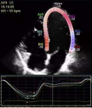
了解GLS 定义前首先让我们看看什么是长轴应变;Longitudinal strain是指对象在某一方向上的相对于初始长度的变化。应变(strain)= (Lt - L0) / L0 %; 其中Lt是在时间t的长度,L0是时间0的初始长度。
最常用的基于应变对LV整体收缩功能测量就是GLS即整体长轴应变,通常通过斑点追踪超声心动图评估。在2DE上,峰值GLS描述了左室舒张末期和收缩末期之间LV心肌的相对长度变化。GLS= (MLs- MLd)/ MLd; 其中ML是收缩末期(MLs)和舒张末期(MLd)的心肌长度,因为MLs小于MLd,所以峰值GLS为负数。当描述应变增加或减少时,GLS的负数表达往往会引起混淆。为此,超声心腔结构测量指南建议所有涉及应变改变的参考文献,在提及应变的增加或减少时,应使用绝对值,以避免混淆。测量时要选取最佳的图像质量、最大的帧频、并将心腔短切的可能性降至最低。 GLS测量应在三个标准心尖切面中进行,取其平均值。测量时,先取心尖长轴切面,利用主动脉瓣开放和关闭的喀喇音,或在M型图像上的主动脉瓣开放和关闭,来确定主动脉瓣的关闭。如果在一个切面上有两个心肌节段局部跟踪不满意,应取消GLS的计算。对于此类病例,可采用其他替代方法估价LV长轴功能,如二尖瓣环收缩期运动幅度,或组织多普勒显像得出的二尖瓣环收缩速度峰值(S’)。 另外由于各超声和软件厂家间差异太大以及年龄和心脏负荷对GLS的影响,建议对同一患者的GLS评估应使用相同厂家的设备和相同软件。测量推荐使用心肌中层(mid wall )GLS的测量值,有庞大的资料证实其临床价值和可重复性。
各种评价左室功能方法介绍及其优缺点比较
线性面积法:
优点:时间分辨率高,光标同轴性差,左心室形态不规则时,数据不可靠。
容量法:
优点: LV形态有一定校正
缺点:双平面有对左心室的评估有一定盲区
整体长轴应变
优点:不依赖于角度的选择,有大量的预后数据
缺点:耗时,对图像质量依赖强,各个厂家提供的参数不同
GLS的应用价值
GLS可以为静息的心功能提供更多对预后的预测,也可以预测瓣膜患者术后左室功能恢复。一项在4172名急性心力衰竭的患者当中进行的里程碑的研究显示无论是在射血分数保留的心衰,还是射血分数降低的心衰中,GLS的测量相对于EF能更好的进行危险分层,GLS改进1%,其病死率下降5%。1另一研究显示在射血分数降低的患者中,即使治疗后EF恢复,GLS仍然存在异常,并且提示其不良预后。2这些研究提示即使EF改进的患者仍然存在左心室功能不全,也需要积极治疗。也有研究证实GLS 可以在心衰stage B的无症状患者中预测将来心衰的发生。19
此外,GLS 的另外一个价值就是对化疗药物心脏毒性的监测,并且也已经写入指南。通过GLS可以早期发现心脏损害,7并及时采取保护措施,相关的大型随机对照研究正在进行当中。10
GLS 在射血分数保留心衰的表型鉴别中也有一定价值。The Framingham 的超声研究显示在过去的30年,HFpEF 的负担加重,其诊断依赖于超声及影像,但超声心动除了能提供EF,左心房的充盈压外,其他信息相对匮乏。通过GLS 还可将HFpEF与左心室肥厚相鉴别,后者对治疗反应更有效果。23
GLS 在瓣膜心脏病中的应用
瓣膜疾病进展有其自然的发展历程,但在某些人群的进展可能会有所不同,需要采取优先的干预策略。目前,指南在瓣膜疾病是否需要干预的决策高度依赖于EF, 但在主动脉和二尖瓣反流方面,GLS似乎可以对治疗策略提供更精细的调整,优于基于EF的决策策略,这两种病变的最佳手术时机可能非常复杂。在两种情况下,GLS都可以进行更好的风险分层和结果估计30-31。
近期,JAMA发表一项研究,旨在分析LV GLS受损和自发LV GLS对于左室射血分数保留(LVEF)患者无症状重度AS的预后判断价值。研究发现与年龄匹配和性别匹配的人群相比,无症状重度AS更容易出现严重LV GLS受损。经过平均12个月的随访期,在LVEF没有显著改善的情况下,患者的LV GLS出现明显恶化(P<0.001)。基线时LV GLS受损的患者出现症状(P=0.02)和需要进行主动脉瓣干预治疗(P=0.03)的危险更高。以LV GLS受损为特征的亚临床心肌功能障碍常见于LVEF保留的无症状重度AS患者。随时间推移,患者的LV GLS进一步恶化。研究显示,基线时LV GLS受损与进展到AS症状期以及需主动脉瓣干预的风险增加具有显著相关性。
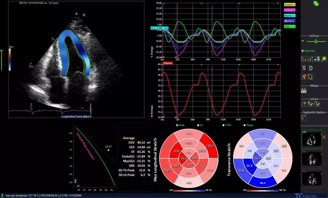
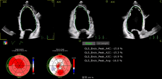
这是一个主动脉瓣重度狭窄患者,EF 63%完全正常,但左室整体应变GLS 15.8%,已经略减低(GLS正常大于16%)
超声心动图的其他进展
LV应变曲线可以用来分析LV的机械离散度,在预测致死性心律不齐的右心室发育不良患者及无心律失常史的其突变阳性家庭成员危及生命的心律失常风险中起一定作用。将来,这可能是一个支持参数,可用于确定该患者是否是植入式心脏复律除颤器的适应证。11
左心房(LA)应变在疾病中的作用:我们越来越了解左心房(LA)在健康和疾病中的作用,LA可以作为左室舒张功能障碍的晴雨表。应变在心房中起着重要的作用,也是衰老和疾病的标志。LA应变是HFrEF和HFpEF心力衰竭的预后因素,远远超过仅由左心室形态学提供的信息,是舒张功能障碍的早期标志。LA应变同时可以帮助预测将来的事件,例如房颤中的卒中,未来的舒张功能障碍指南可能会纳入LA数据,以对该人群的诊断和预后亚组进行分类和分层。12-13
高帧频成像(High frame Rate): 高帧频成像是HFpEF 超声影像学诊断最大的进展之一,可以直接对心肌僵硬度进行测量,可以通过脉冲后的自然剪切测量心肌的僵硬度从而判定HCM 僵硬度恶化, 高帧频成像还可以用来无创评估冠脉血管、LV心腔血流模式以及对心肌血流进行定量测定。24-26
此外三维、四维超声心动图以及腔内超声心动图对瓣膜的介入治疗指导也有着更大的价值,我们将在后面的栏目中深入介绍
总之,希望心内科医生能早日带上超声的翅膀去飞翔。
参考文献:
1.Park J.J., Park J.-B., Park J.-H., Cho G.-Y. (2018) Global longitudinal strain to predict mortality in patients with acute heart failure. J Am Coll Cardiol 71:1947–1957.FREE Full TextGoogle Scholar
2.Merken J., Brunner-La Rocca H.-P., Weerts J., et al. (2018) Heart failure with recovered ejection fraction. J Am Coll Cardiol 72:1557–1558.FREE Full TextGoogle Scholar
3.Mirea O., Pagourelias E.D., Duchenne J., et al. (2018) Variability and reproducibility of segmental longitudinal strain measurement: a report from the EACVI-ASE Strain Standardization Task Force. J Am Coll Cardiol Img 11:15–24.Google Scholar
4.Mirea O., Pagourelias E.D., Duchenne J., et al. (2018) Intervendor differences in the accuracy of detecting regional functional abnormalities: a report from the EACVI-ASE Strain Standardization Task Force. J Am Coll Cardiol Img 11:25–34.Google Scholar
5.Thavendiranathan P., Negishi T., Coté M.A. (2018) Single versus standard multiview assessment of global longitudinal strain for the diagnosis of cardiotoxicity during cancer therapy. J Am Coll Cardiol Img 11:1109–1118.Google Scholar
6.Medvedofsky D., Maffessanti F., Weinert L., et al. (2018) 2D and 3D echocardiography-derived indices of left ventricular function and shape: relationship with mortality. J Am Coll Cardiol Img 11:1569–1579.Google Scholar
7.Henry M.L., Niu J., Zhang N. (2018) Cardiotoxicity and cardiac monitoring among chemotherapy-treated breast cancer patients. J Am Coll Cardiol Img 11:1084–1093.Google Scholar
8.Nolan M.T., Marwick T.H., Plana J.C., et al. (2018) Effect of traditional heart failure risk factors>↵Avila M.S., Ayub-Ferreira S.M., de Barros Wanderley M.R. Jr.., et al. (2018) Carvedilol for prevention of chemotherapy-related cardiotoxicity: the CECCY Trial. J Am Coll Cardiol 71:2281–2290.FREE Full TextGoogle Scholar
9.Negishi T., Thavendiranathan P., Negishi K., et al. (2018) Rationale and design of the strain surveillance of chemotherapy for improving cardiovascular outcomes. J Am Coll Cardiol Img 11:1098–1105.Google Scholar
10.Lie Ø.H., Rootwelt-Norberg C., Dejgaard L.A., et al. (2018) Prediction of life-threatening ventricular arrhythmia in patients with arrhythmogenic cardiomyopathy. J Am Coll Cardiol Img 11:1377–1386.Google Scholar
11.Morris D.A., Belyavskiy E., Aravind-Kumar R., et al. (2018) Potential usefulness and clinical relevance of adding left atrial strain to left atrial volume index in the detection of left ventricular diastolic dysfunction. J Am Coll Cardiol Img 11:1405–1415.Google Scholar
12.Pathan F., Sivaraj E., Negishi K., et al. (2018) Use of atrial strain to predict atrial fibrillation after cerebral ischemia. J Am Coll Cardiol Img 11:1557–1565.Google Scholar
13.Lupón J., Gavidia-Bovadilla G., Ferrer E., et al. (2018) dynamic trajectories of left ventricular ejection fraction in heart failure. J Am Coll Cardiol 72:591–601.FREE Full TextGoogle Scholar
14.Sze E., Samad Z., Dunning A., et al. (2018) Impaired recovery of left ventricular function in patients with cardiomyopathy and left bundle branch block. J Am Coll Cardiol 71:306–317.FREE Full TextGoogle Scholar
15.Huckstep O.J., Williamson W., Telles F., et al. (2018) Physiological stress elicits impaired left ventricular function in preterm-born adults. J Am Coll Cardiol 71:1347–1356.FREE Full TextGoogle Scholar
16.Guzzardi M.A., Liistro T., Gargani L., et al. (2018) Maternal obesity and cardiac development in the offspring. J Am Coll Cardiol Img 11:1750–1755.Google Scholar
17.Vasan R.S., Xanthakis V., Lyass A. (2018) Epidemiology of 3 decades. left ventricular systolic dysfunction and heart failure in the Framingham study: an echocardiographic study over. J Am Coll Cardiol Img 11:1–11.Google Scholar
18.Wang Y., Yang H., Huynh Q., Nolan M., Negishi K., Marwick T.H. (2018) Diagnosis of nonischemic stage B heart failure in type 2 diabetes mellitus: optimal parameters for prediction of heart failure. J Am Coll Cardiol Img 11:1390–1400.Google Scholar
19.Kosmala W., Przewlocka-Kosmala M., Rojek A., et al. (2018) Association of abnormal left ventricular functional reserve with outcome in heart failure with preserved ejection fraction. J Am Coll Cardiol Img 11:1737–1746.Google Scholar
20.Selmeryd J., Henriksen E., Dalen H., Hedberg P. (2018) Derivation and evaluation of age-specific multivariate reference regions to aid in identification of abnormal filling patterns. J Am Coll Cardiol Img 11:400–408.Google Scholar
21.Omar A.M., Shameer K., Narula S., et al. (2018) Artificial intelligence-based assessment of left ventricular filling pressures from 2-dimensional cardiac ultrasound images. J Am Coll Cardiol Img 11:509–510.Google Scholar
22.Mordi I.R., Singh S., Rudd A., Srinivasan J., Frenneaux M., Tzemos N., Dawson D.K. (2018) Comprehensive echocardiographic and cardiac magnetic resonance evaluation differentiates among heart failure with preserved ejection fraction patients, hypertensive patients, and healthy control subjects. J Am Coll Cardiol Img 11:577–585.Google Scholar
23.Maresca D., Correia M., Villemain O., et al. (2018) Noninvasive imaging of the coronary vasculature using ultrafast ultrasound. J Am Coll Cardiol Img 11:798–808.Google Scholar
24.Toulemonde M.E.G., Corbett R., Papadopoulou V., et al. (2018) High frame-rate contrast echocardiography: in-human demonstration. J Am Coll Cardiol Img 11:923–924.Google Scholar
25.Villemain O., Correia M., Khraiche D., et al. (2018) Myocardial stiffness assessment using shear wave imaging in pediatric hypertrophic cardiomyopathy. J Am Coll Cardiol Img 11:779–781.Google Scholar
26.Park C.S., Park J.B., Kim Y., et al. (2018) Left ventricular geometry determines prognosis and reverse J-shaped relation between blood pressure and mortality in ischemic stroke patients. J Am Coll Cardiol Img 11:373–382.Google Scholar
27.Donnellan E., Griffin B.P., Johnston D.R., et al. (2018) Rate of progression of aortic stenosis and its impact>↵Zilberszac R., Heinze G., Binder T., Laufer G., Gabriel H., Rosenhek R. (2018) Long-term outcome of active surveillance in severe but asymptomatic primary mitral regurgitation. J Am Coll Cardiol Img 11:1213–1221.Google Scholar
28.Alashi A., Mentias A., Abdallah A. (2018) Incremental prognostic utility of left ventricular global longitudinal strain in asymptomatic patients with significant chronic aortic regurgitation and preserved left ventricular ejection fraction. J Am Coll Cardiol Img 11:673–2.Google Scholar
29.Kim H.M., Cho G.Y., Hwang I.C., et al. (2018) Myocardial strain in prediction of outcomes after surgery for severe mitral regurgitation. J Am Coll Cardiol Img 11:1235–1244.Google Scholar
30.Dejgaard L.A., Skjølsvik E.T., Lie O.H., et al. (2018) The mitral annulus disjunction arrhythmic syndrome. J Am Coll Cardiol 72:1600–1609.FREE Full TextGoogle Scholar
31.Topilsky Y., Maltais S., Medina Inojosa J., et al. (2019) Burden of tricuspid regurgitation in patients diagnosed in the community setting. J Am Coll Cardiol Img 12:433–442.Google Scholar
32.Treibel T.A., Kozor R., Fontana M., et al. (2018) Sex dimorphism in the myocardial response to aortic stenosis. J Am Coll Cardiol Img 11:962–973.Google Scholar
33.Treibel T.A., Kozor R., Schofield R., et al. (2018) Reverse myocardial remodeling following valve replacement in patients with aortic stenosis. J Am Coll Cardiol 71:860–871.FREE Full TextGoogle Scholar
34.Goublaire C., Melissopoulou M., Lobo D., et al. (2018) Prognostic value of exercise-stress echocardiography in asymptomatic patients with aortic valve stenosis. J Am Coll Cardiol Img 11:787–795.Google Scholar
35.Annabi M.S., Touboul E., Dahou A. (2018) Dobutamine stress echocardiography for management of low-flow, low-gradient aortic stenosis. J Am Coll Cardiol 71:475–485.FREE Full TextGoogle Scholar
36.Dahou A., Clavel M.A., Capoulade R., et al. (2018) B-type natriuretic peptide and high-sensitivity cardiac troponin for risk stratification in low-flow, low-gradient aortic stenosis: a substudy of the TOPAS study. J Am Coll Cardiol Img 11:939–947.Google Scholar
37.Kataoka A., Watanabe Y., Kozuma K. (2018) Prognostic impact of low-flow severe aortic stenosis in small-body patients undergoing TAVR: the OCEAN-TAVI registry. J Am Coll Cardiol Img 11:659–669.Google Scholar
38.Hahn R.T., Leipsic J., Douglas P.S., et al. (2019) Comprehensive echocardiographic assessment of normal transcatheter valve function. J Am Coll Cardiol Img 12:25–34.Google Scholar
39.Lee P.H., Song J.K., Kim J.S., et al. (2018) Cryptogenic stroke and high-risk patent foramen ovale: the DEFENSE-PFO trial. J Am Coll Cardiol 71:2335–2342.FREE Full TextGoogle Scholar
40.Fauchier L., Cinaud A., Brigadeau F., et al. (2018) Device-related thrombosis after percutaneous left atrial appendage occlusion for atrial fibrillation. J Am Coll Cardiol 71:1528–1536.FREE Full TextGoogle Scholar
41.Salaun E., Sportouch L., Barral P.-A., et al. (2018) Diagnosis of infective endocarditis after TAVR. J Am Coll Cardiol Img 11:143–146.Google Scholar
42.Egbe A., Pislaru S.V., Ali M.A., et al. (2018) Early prosthetic valve dysfunction due to bioprosthetic valve thrombosis: the role of echocardiography. J Am Coll Cardiol Img 11:951–958.Google Scholar
43.Bhavnani S.P., Sola S., Adams D., et al. (2018) A randomized trial of pocket-echocardiography integrated mobile health device assessments in modern structural heart disease clinics. J Am Coll Cardiol Img 11:546–557.Google Scholar